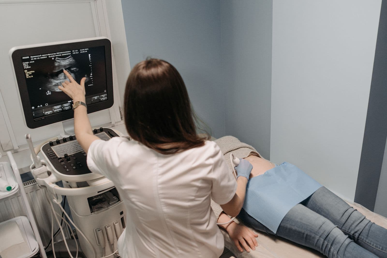

Screening Is Not One-Size-Fits-All
For decades, breast cancer screening advice boiled down to a single message: get a mammogram. That is still excellent advice — mammography saves lives — but it is not the whole story. A truly good screening plan accounts for your personal risk, your breast density, and your family history. For a meaningful number of women, a standard mammogram alone is not enough.
Understanding why, and when to layer in additional tools, is one of the most important conversations you can have with your primary care physician.
The Baseline: What Standard Screening Looks Like
The U.S. Preventive Services Task Force now recommends biennial screening mammography for women ages 40 to 74. That is a meaningful shift from the older "start at 50" recommendation, and it aligns more closely with what the American Cancer Society and CDC have been saying for years.
For women at average risk, that biennial schedule is a reasonable baseline — and many clinicians, myself included, prefer annual mammograms from 40 onward, especially in women with any additional risk factors. Routine screening fits naturally into the rhythm of preventative care in St. Pete and the annual physical.
Digital Breast Tomosynthesis (3D Mammography)
If your imaging center offers it, tomosynthesis is generally preferred over 2D mammography alone. It captures thin slices of the breast, which improves cancer detection and reduces false-positive callbacks — especially helpful in younger women and those with denser tissue.
The Dense Breast Problem
Roughly 40 percent of women have dense breast tissue on mammography. Dense tissue matters for two reasons:
- It independently raises breast cancer risk.
- It reduces the sensitivity of mammography — dense fibroglandular tissue and cancer both appear white on the image, which can hide tumors.
FDA rules now require that mammogram reports notify you of your breast density. If your report mentions "heterogeneously dense" or "extremely dense" tissue (categories C and D), that is your cue to have a conversation about supplemental screening.
Options to consider alongside mammography in dense breasts include:
- Screening breast ultrasound. Useful supplement for women with dense breasts at average risk. It catches some cancers mammography misses, at the cost of more false positives.
- MRI. Reserved for higher-risk women — more on this below.
- Contrast-enhanced mammography, where available.
When MRI Becomes Part of the Plan
Breast MRI is the most sensitive imaging test we have, but it is expensive and produces more false positives than mammography, so we use it selectively. Current guidance from American Cancer Society and major oncology bodies supports annual MRI in addition to mammography for women with:
- A known BRCA1 or BRCA2 mutation, or a first-degree relative with one
- A lifetime breast cancer risk of 20 percent or greater, calculated using a validated model such as Tyrer-Cuzick
- A history of chest radiation between ages 10 and 30 (for example, after Hodgkin lymphoma treatment)
- Certain other high-penetrance gene mutations (TP53, PTEN, PALB2, CDH1)
- A history of atypical hyperplasia or lobular carcinoma in situ, in some cases
If you have multiple first- or second-degree relatives with breast, ovarian, pancreatic, or prostate cancer — particularly at young ages — a formal risk calculation belongs in your chart.
Genetic Testing: Who, and When
Genetic testing has moved from a specialty rarity to a mainstream tool. You should discuss referral for genetic counseling if any of the following apply:
- Breast cancer diagnosed before age 50 in a close relative
- Ovarian cancer at any age in the family
- A male relative with breast cancer
- Multiple breast cancers in one person, or bilateral disease
- Ashkenazi Jewish ancestry plus a family history of breast, ovarian, or pancreatic cancer
- A known pathogenic variant in the family
- Triple-negative breast cancer diagnosed before age 60
A positive result changes screening, sometimes changes surgical considerations, and has implications for family members who may wish to be tested themselves.
Self-Awareness, Not Self-Exams
The formal recommendation for structured monthly breast self-examination has been set aside. The evidence did not show a mortality benefit, and it generated a lot of unnecessary biopsies. What has replaced it is breast self-awareness — knowing how your breasts normally look and feel, and reporting new changes promptly. Lumps, new nipple discharge, skin dimpling, nipple retraction, persistent pain in one spot, or asymmetric changes all warrant a same-week call to your physician.
The same principle applies to other skin and tissue changes — our guide on skin cancer screening in St. Petersburg explains the self-awareness approach for another common cancer.
A Brief Note on Men
Male breast cancer accounts for roughly 1 percent of all breast cancers. Men with a strong family history, a known BRCA2 mutation, or Klinefelter syndrome should be aware of the signs and discuss screening with their physician — routine imaging is not standard, but physical exam and prompt evaluation of any lump are essential.
What Else Moves Your Risk, and What to Do About It
Family history and density get the attention, but several everyday factors shift breast cancer risk enough to change a screening conversation, and a few of them are in your control.
Hormone therapy, weight, and alcohol
Combined estrogen-plus-progestin therapy for menopausal symptoms modestly raises breast cancer risk with longer use, while estrogen alone in women without a uterus does not appear to; our article on menopause hormone therapy risks and benefits walks through that trade-off, which is different for every woman going through menopause. After menopause, obesity raises risk because fat tissue becomes the main source of estrogen, and alcohol raises it in proportion to the amount, with no clearly safe floor. Regular physical activity lowers it. None of these replaces screening, but they are the reason I ask about all three at the visit where we set your schedule.
Medication to lower risk in high-risk women
For women whose calculated risk is high, whether from family history, prior atypical biopsy, or a genetic result, tamoxifen before menopause and raloxifene after it reduce the chance of developing estrogen-receptor-positive breast cancer. They are not for average-risk women, because they carry clot and, for tamoxifen, uterine risks. The USPSTF reviews who benefits, and our BRCA genetic testing article covers the testing that often precedes that decision.
When screening stops earning its keep
Mammography is generally continued while a woman is in good health with a life expectancy of ten years or more, which for many patients here means well past 75. It stops making sense when other illness is more likely to shorten life than a screen-detected cancer, and that is a conversation rather than a birthday. The National Cancer Institute breast screening summary lays out the evidence in plain language.
Building Your Personal Plan
Breast cancer screening works best as a conversation, not a checkbox. I want to know your family history, your breast density, your reproductive history, and any prior biopsies before I recommend a schedule. For some women that means biennial mammography. For others it means annual 3D mammography plus ultrasound, or annual alternating mammography and MRI.
If you are not sure whether your current plan matches your risk, schedule a visit. We can review your history, calculate your lifetime risk, coordinate any additional imaging through our lab and imaging partners, and refer for genetic counseling if it is warranted. Screening done right is one of the highest-value things we do in primary care.


